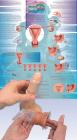
Class pack refill, Classroom-sized refill access to 1 digital interactive case with up to 1 teacher and 30 single-use student accounts. Engaging web-based lab activity helps students learn and apply key forensic science concepts while solving case of this historic double murder
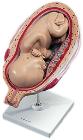
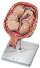
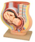
Model, Fetus, month 7, The model shows a fetus in the seventh month in the uterus, All important anatomical structures are labeled, Dimensions: 15 x 32 x 27 cm, weight: 0.86 kg
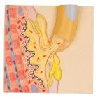
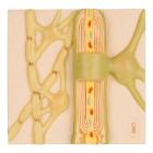
Model Motor End Plate, depicts the neuromuscular junction with striated muscle fiber, The motor end plate of the human nervous system is depicted in colorful anatomical detail, great addition to any lesson on the human nervous, Dimensions : 12 x 11.5 x 3.2 cm
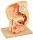
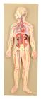
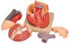
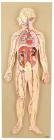
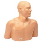
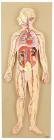
This half-size relief model helps in understand the circulation of blood in body through brain, heart, lung, liver, spleen, kidneys and partial skeleton. Mounted on base. Numbered with key card.
Model Neuron Cell Body, depicts a part of the human nervous system in colorful anatomical detail, neuron model features a typical neuron body with cell organelles, for example mitochondria and many other characteristics, Weight: 0.47 kg, Dimensions : 12.2 x 11.7 x 6.2 cm
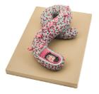
Model, 5th Month Twin Fetuses - Normal Position, shown in a natural position within the uterus, They can be removed for a closer inspection of the uterus, this high quality model is a great tool for studying the anatomy of human development, weight: 0.66 kg
Model Ebola Virus 10000X, represents the virus magnified approximately 100000 times in its unique shape, surface shows the viral membrane with Glycoproteins, A cut-away at one end exposes internal structures major and minor matrix proteins polymerase protein and Ebola RNA
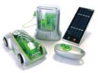
Heat-it Kit, Group, lets you grow and engineer bacteria with a DNA plasmid that can be controlled with heat. Decide when you want the DNA program to turn on by choosing the temperature you grow the bacteria at, to create tree ring pat
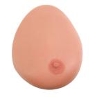
Model Single Breast, With Benign Tumor, Dimensions: 25.4 x 17.8 x 17.8 cm, made of 3B SKINlike* silicone with simulated benign tumour for the demonstration of ultrasonic B-image mode with Ultrasonic Echoscope GS200
Model Myelin Sheaths Of The CNS, shows the glial cells which build the insulating layer around the axons of the central nervous system, great addition to any lesson about the human nervous system, Weight: 0.4 kg, Dimensions : 12.2 x 11.7 x 3.6 cm
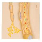
Model Schwann Cells Of The PNS, depicts a Schwann cell with sectioned core, depicts the schwann cell in colorful anatomical detail, great addition to any lesson on the human nervous system, Weight: 0.4 kg, Dimensions : 12.2 x 11.7 x 3.2 cm
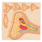
Model Synapse, features the endoplasmic reticulum, mitochondria and the membranes of the synaptic gap, Also depicted by the synapses model are 5 smaller relief models of the main synapse variations, great addition to any anatomical lesson, Dimensions : 12 x 11.5 x 2.7 cm
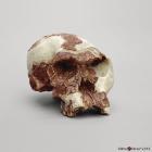
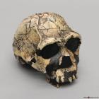
Model Homo Habilis Oh 24 Cranium, 1.8 MYA, was discovered by P. Nzube in 1968. This nearly complete but very badly crushed specimen constituted the oldest hominid found in Olduvai Gorge, Tanzania, Size: 18.3L x 11.6W x 10.1H (cm)
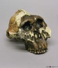
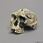
Model H. Ergaster Knm-Er 3733 Cranium, 1.75 MYA, with dentition was discovered by B. Ngeneo in 1975 in Koobi Fora, Kenya. Several teeth are intact, but no mandible was ever found, Size: 19.1L x 13W x 13.5H (cm)
Model A. Boisei Knm-Er 406 Cranium, 1.7 MYA, was discovered by R. Leakey at Kenya, in 1969. This discovery helped support the classification of boisei as a separate species of Australopithecus, Size: 19.9L x 17.1W x 11.8H (cm)
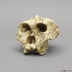
Model A.Africanus Sts 5 Mrs. Ples Cranium, 2.5 MYA, was discovered in 1947 by R. Broom and J. Robinson in Sterkfontein, Transvaal, South Africa, Size: 17.2L x 12.3W X 13.1H (cm)
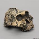
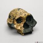
Model A. Boiseioh5(Zinjanthropus)Cranium, 1.8 MYA, most famous fossil from Olduvai Gorge, Tanzania. OH5 was discovered by Mary Leakey in 1959. The accepted genus name has since changed to Australopithecus, Size: 19.5L x 16.1W X 17.2H (cm)
Model Australopithecus Boisei Femaleknm Er 732 Cranium, 1.7 MYA. Female, was discovered in 1970 at Koobi Fora, Kenya by R. Leakey and H. Mutua and described in Nature in 1971, Size: 16.6L x 14.1W x 10.2H (cm)
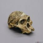
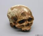
Model Homo Habilis Knm-Er 1813 Cranium, 1.9 MYA, was discovered by K. Kimeu in 1973 at Koobi Fora, Kenya, and described by R. Leakey in Nature in 1973, Size: 17.1L x 11.8W x 11.6H (cm)
Model A. Robustus Sk-48 Cranium, 1.5-2 MYA, was discovered by Fourie in South Africa in 1950. SK-48, formerly Paranthropus crassidens, greatly increased what is known about australopithecines, Size: 15.6L x 15.1W X 12.3H (cm)
Model Homo Sapiens Cro-Magnon 1 Cranium, 30,000-32,000 YA, discovered by L. Lartet and H. Christy on a cliff in 1868 (during the construction of railway lines in Les-Eyzies, France), Size: 21L x 15.2W X 17H (cm)
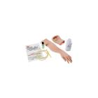
simulator, Amniotic fluid insert, for the birth simulator, P90 Pro, can be used for practicing on vertical amniotic sacs, It contains an additional pouch of 250 ml of artificial amniotic fluid and 100 amnion inserts, Dimesnions: 15 x 6 x 16 cm, Weight: 0.9 kg
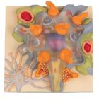
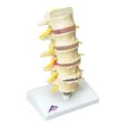
Model Stages Of Disc Prolapse, includes: Intervertebral disc in healthy condition, Lumbar vertebra (L1) without degenerative changes. Intervertebral disc with protrusion, Lumbar vertebra (L2) with beginning degenerative changes, Dimensions : 22 cm