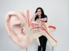
Model, World's Largest Ear, 15 times full-size, 3 part, at 15 times life-size, this 3-part human ear is suitable for museums and special collections, The giant ear represents the outer, middle and inner ear, Dimensions: 130 x 120 x 60 cm, weight: 72 kg
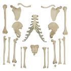
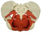
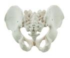
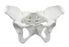
It Illustrate the morphological distinctions between male and female pelvic structures. Each pelvis includes the left and right innominates with pubic symphysis, 4th and 5th lumbar vertebrae with intervertebral discs, the sacrum and coccyx.
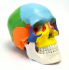
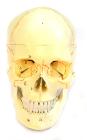
Each part on the skull is numbered. Jaw is spring loaded. Skull cap is removable for further interior inspection. Included with the model is a sheet with each numbers name for identification.
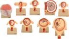
Set of nine models, showing the following stages. 1. Embryo 6 days old 2. 1st month of gestation. 3. Uterus with embryo in 3rd month of gestation.4. Uterus with fetus, in 4th month. 5. Uterus with fetus, placenta and umbilical cord.6. 5th month. 7. 7th month pregnancy.
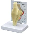
Model of human elbow joint. Mounted on base. Features upper half of radius and ulna, lower half of humerus and the ligaments of the elbow joint, to show hinge joint, Numbered with English Key Card, Size 17x 22 x 22 cm approx, Weight 250 g
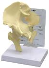
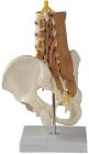
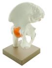
Model of human hip joint. With simulated pubofemoral ligament, Mounted on base, represents the upper femur and pelvic bone and the way that they move, Numbered with English Key Card, Size 22 x 12 x 25 cm approx. Weight 800 g approx
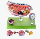
Model of the Ovary, Enlarged approximately 10 times, in SOMSO-Plast* Horizontal section parallel to the mesovarian margin with presentation of the follicles in different maturation phases, corpus rubrum, luteum and albicans, Separates into 13 parts. Mounted
Illustrate the morphological distinctions between male and female pelvic structures. Each pelvis includes the left and right innominates with pubic symphysis, 4th and 5th lumbar vertebrae with intervertebral discs, the sacrum and coccyx.