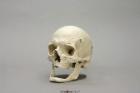
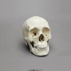
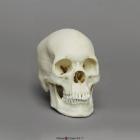
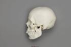
Model Human Elderly European Male Skull (80 years old), Features of advanced age are limited to marked extra & intracranial suture closure, irregular bony exostosis & further suggested by edentulous nature of jaws, Size: 20.4x13.7x17.6cm
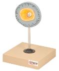
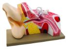
Model shows both sides of an eye, enlarged 5 x. One side of the model shows the eye socket with a sagittal cutaway and the background to the eye and the electron microscopic fine structure of the retina are shown separately.
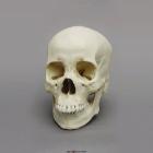
Model Human Female American Indian Skull, Includes broad, flattened, forward-projecting zygomas, facial bones are vertically aligned with shallow nasal depression, moderate nasal spine, orthognathic jaw & vertical chin, Size: 18.5x14x18.8cm
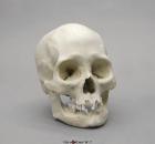
Model Human Female Asian Economy Skull, Features no cranial sites for musculofascial attachment, a narrow ascending mandibular ramus, smooth nasion, sharp supraorbital margins & a rounded inferior border of mandible, Size: 17.1x12x16.5cm
Model Human Female Asian Skull, include a flat nasal root, an obtuse nasal angle, a somewhat rounded dental arcade in the upper jaw, mild to moderate prognathism and shovel-like incisors in the upper jaw, Size: 19.4L x 12.9W X 17.3H (cm)
Model Human Female Asian Skull, include moderate alveolar prognathism, flat nasal region, round/square anterior nasal aperture, lack of a nasal sill, no nasal gutter, round maxillary arcade, and short nasal spine, Size: 20.5L x 12.3W X 18.7H (cm)
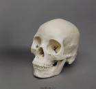
Model Human Female European Skull, generally gracile, specimen is not definitively female, serve as a good discussion piece in a classroom setting for the diagnostic limitations in the determination of sex, Size: 19.2L x 12.6W x 19H (cm)
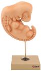
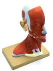
Life size model showing the outer superficial muscles, vessels, nerves and head with muscles on one side. On the outer side details of median section such as brain, mouth, larynx are shown. Includes key card.
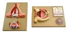
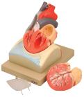
Enlarged, sectioned so that both ventricles and atria open to expose the valves. Large blood vessels near the heart and musculature of the heart are shown. Separates into 4 parts. On base.
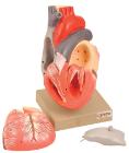
Life size model dissectible in 2 parts. The anterior heart wall can be removed to show the left and right ventricles and atria as well as the tricuspid, pulmonary, mitral and aortic valves. Mounted on base.
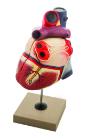
Sectioned through the ventricles and auricles. The bicuspid and tricuspid semilunar and sigmoid valves are shown. Separates into 3 parts. Mounted on base
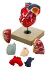
Enlarged. Sectioned so that both ventricles and atria open to expose the valves. Large blood vessels near the heart and musculature of the heart are shown. Separates into 7 parts.